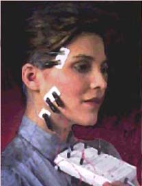


Computerized
Electro-diagnostic Instrumentation
ADVANCED TECHNIQUES FOR PRECISION AND
ACCURACY
COMPUTERIZED ELECTRO-DIAGNOSTIC INSTRUMENTATION -
An Advanced Technology


Invented by Dr. Bernard Jankelson and further developed and perfected by
his son Dr. Robert R. Jankelson (Myotronics/ Normed, Inc. Tukwila,
Washington).
A variety of other techniques have been developed to diagnose TMJ diseases and
disorders including mandibular jaw tracking, surface electromyography and
sonography. The use of computerized electro-diagnostics is a more sophisticated
approach to accurately and objectively define and treat TMJ/ TMD. In the past
these technologies were not available thus resulting in erroneous
conclusion, misdiagnosis, and misguided treatments.
Although there are some opponents that say that there is inadequate evidence to
support the use and effectiveness of such diagnostics instrumentation, it is
clear that they are misinformed and do not understand how the instrumentation
can be used and implemented to aid in the diagnosis and treatment of TMJ.
Computerized Mandibular Scanning is a more complex assessment of
mandibular function using biomedical instrumentation which measures
 the rotational movement in the frontal and sagittal planes thus confirming a
neuromuscular dysfunction. The computerized mandibular scanner measures jaw
movement (both qualitatively and quantitatively in several dimensions) to
within 0.1 millimeters of accuracy. With a magnetic tracking device and sensor
array, it projects the data on a calibrated computer monitor.
The CMS measures jaw movement is far more accurate than the eye, making it
possible to document characteristics of mandibular motion considered
significant to evaluate jaw function. It also identifies the amount of free
space, the swallowing pattern, and the quality of the occlusion, and
substantiates the presence of disc derangements and their prognosis for
reduction. It is a multi-dimensional assessment of torquing movements used to
differentiate between contributing factors of a pathologic position to a
non-pathologic position on opening and closing of the mandible. It is used in
conjunction with EMG recordings.
Graphic recording of opening/ closing paths of jaw movements from the side and
front views can be analysed to assess abnormal mandibular paths of movement.
The speed at which the jaw can open and close is also simultaneously recorded.


Sagital/
Frontal views of jaw movement
Range
of Motion can be measured accurately
The literature supports the
efficacy of mandibular tracking in the diagnosis and treatment of TMJ/ MSD.
There are over 22 controlled published studies that further support the
rationale for mandibular jaw tracking.
There are 25 additional supporting referenced studies confirming the same.
There are numerous other studies that document the clinical efficacy and
validity of computerized mandibular scanning.
Surface electromyography is a series of tests to more specifically delineate
and define hypertonic musculature in the compromised TMJ patient.  These series of tests are necessary to differentially diagnose between
intra-capsular interference (mensical or otherwise) and extra-capsular
interference (influence of the surrounding hypertonic muscular matrix) so as
to determine the predominant dysfunctions. Surface electrodes are placed
over the muscles which in turn send impulses to the recording instrument.
Defining the etiology of the TMJ patient's predominate neuromuscular
dysfunctions will preclude misdirected palliative treatment regimens.
Surface electromyography (EMG) utilizes eight channels monitoring the right
and left posterior temporalis muscles, right and left anterior temporalis
muscles, right and left masseters, and right and left anterior digastric
muscles. A clinical hands-on muscle palpation examination is not able to
quantify and objectively record muscle hypertonicity with out subjective
intervention.
Muscles of the face and jaw can be recorded to determine hyperactive muscle
activity and/ or resting muscle activity. A strained jaw position can effect
muscle activity. The objective is to determine the optimal resting jaw
position at physiologic rest that harmonizes with resting EMG levels.


Hyperactive/
Strained Muscles
Calm/
Rested Muscles
There is a broad body of literature that supports the physiologic basis for
using surface EMG as an aid in assessment of muscle function/ dysfunction.
(38 + studies support this ending with Lynn et al, 1992).
There is substantial evidence based upon controlled studies that confirm
that surface EMG is reliable and reproducible. (18 studies ending with Dean
et al., 1992).
87 studies verifying the use, safety, and efficacy of EMG to monitor
masticatory muscle function/ dysfunction.
"In summary, based on well controlled empirical and clinical studies
that have been conducted in several universities over the past three decades
throughout the world, there is unequivocal evidence to strongly support the
use of EMG for the evaluation and diagnosis of temporomandibular
disorders." - Robert Jenkelson, D.D.S.
Sonography utilizes a kinesograph to measure intracapsular TMJoint sounds
against normalized data,  duration of these sounds, exact location of the occurrence of these sounds
during jaw opening/ closing, or lateral excursions, and a spectral frequency
analysis of the sound. Without this information, one could not restore
function free of intracapsular interference resulting in decreased muscle
tenderness on palpation, an increased range of motion free of restrictions
and resolve patient complaints of pain). A pair of ultrasensative
transducers are held in place by a lightweight headset over the
temporomandibular joints. Vibrations from each joint during opening and
closing of the mandible are monitored by the transducers, amplified and
inputted into a computer for display, analysis and data storage. The joint
sounds are analy
Sound vibration recordings when the jaw is opened and closed.


Joint
Pathology
Normal
Quite Joint
Transcutaneous electrical nerve stimulation is a specific therapy for the
treatment and resolution of pain related to
Although the use of TENS is a mode of treatment it can be used most
effectively when used in conjunction with CMS and EMG recordings
simultaneously in objectively documenting and diagnostically gathering
information before, during and after treatment.
The efficacy of low frequency
TENS in the diagnosis and treatment of TMJ/ MSD has been clearly confirmed in
the published literature. It is clear and unequivocal that low frequency TENS
(.05 Hz - 10 Hz) is both safe and efficacious for muscle relaxation and pain
control. It is clear that low frequency TENS has a high degree of specificity
when utilized for craniofacial pain.
(Over 44 internationally published studies support and confirm this fact).
There is more than adequate
confirming evidence to support the effectiveness of such diagnostic
instrumentation as verified and confirmed by the American Dental Association
(ADA) and the Food and Drug Administration (FDA).
The American Dental
Association's Council on Scientific Affairs
has awarded surface electromyography (SEMG), Computer Mandibular Scanning
(CMS), and Sonography its "Seal of Acceptance", as diagnostic
aids in the management of temporomandibular disorders.
(Report on Acceptance of TMD
Devices, ADA Council on Scientific Affairs, JADA, Vol. 127, November 1996)
The U.S. Food and Drug
Administration has
granted 510k status to each of these mentioned devices for use in the
diagnosis and management of TMD in my practice.
This reflects that the U.S. Government and the dental profession acknowledges
the safety and efficacy of the devices as recording and measuring devices used
in the diagnosis and management of TMD and orofacial pain.
Go to: MYOTRONICS/NOROMED
for more about this high technology company.
They can also be reached at:
15425 - 53rd Ave
S Tukwila, WA 98188
(206) 243-4214 - Fax: (206) 243-3625
info@myotronics.com